

The 2026 Nobel Prize in Physiology or Medicine went to Karl Deisseroth, Peter Hegemann and Georg Nagel on October 5, for discoveries in light-gated ion channels and optogenetics [1]. Their original work began with a basic question: Why do green algae move toward light?
Optogenetics gives researchers a capability that has been sought for decades: the ability to control the activity of genetically defined nerve cells with light. The approach makes it possible to manipulate neural activity with exceptional spatial and temporal precision and investigate how specific cells and circuits contribute to memory, emotion, movement, and behavior.
For neuroscience researchers, the Nobel Prize recognizes not only a remarkable biological discovery, but also the development of a technology that continues to shape how the brain is studied today.
From channelrhodopsins to control of neural circuits
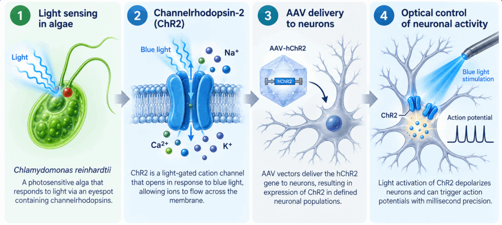
The origins of optogenetics can be traced to studies of the light-sensitive green alga Chlamydomonas reinhardtii. Work by Peter Hegemann, Georg Nagel, and colleagues led to the identification of channelrhodopsins, including channelrhodopsin-2 (ChR2), a directly light-gated cation channel that converts light exposure into ion flow across the cell membrane [2].
Karl Deisseroth and colleagues subsequently demonstrated that ChR2 could be expressed in mammalian neurons and used to trigger action potentials with millisecond-scale precision using blue light [3]. The approach was soon extended to intact neural circuits using viral gene delivery and optical fibers. In an early in vivo demonstration, light activation of ChR2-expressing neurons in the mouse motor cortex produced measurable whisker movement [4] (Figure 1).
This ability to manipulate genetically defined neurons with precise timing changed how researchers could study neural circuits. Rather than only correlating neuronal activity with a physiological or behavioral response, optogenetics made it possible to directly test whether activity in a selected neuronal population was sufficient or necessary for that response [5].
AAV opsin vectors as tools for in vivo optogenetics
The discovery of channelrhodopsins provided the molecular switch, but effective neuroscience experiments also require a way to deliver that switch to the appropriate cells. Viral gene delivery was central to moving optogenetics from cultured neurons into intact tissues. Adeno-associated virus (AAV) vectors were used early to express ChR2 in retinal ganglion cells in vivo [6] and this technique has since become an important platform for expressing optogenetic constructs in defined neuronal populations.
The scientific background accompanying the Nobel Prize specifically highlights the extension of ChR2 experiments to intact brain tissue through viral delivery [1]. Later work used the same route to restrict expression and define neuronal populations.
For researchers designing these experiments, the opsin itself is only one part of the construct. Promoter selection, capsid serotype, reporter choice, and genetic targeting strategy can all impact where the construct is expressed and how easily expression can be monitored. This creates a need for optogenetic vectors that can be matched to the biological question, rather than relying on a single universal configuration.
Choosing an AAV opsin vector and controls
The original ChR2 established the principle of light-driven neuronal activation, but subsequent engineering produced variants with different photocurrent amplitudes, channel kinetics, and light sensitivities. These properties involve trade-offs: variants optimized for faster closing may produce smaller photocurrents, whereas variants with prolonged open states can increase light sensitivity at the expense of precise spike-by-spike control. The appropriate ChR2 variant therefore depends on the experimental objective [7-10].
Driving activity answers half of the question while silencing answers the other half. Light-driven ion pumps such as enhanced halorhodopsin (eNpHR3.0) move chloride into the cell under yellow light and hyperpolarize it, with currents stable over prolonged illumination [11]. Archaerhodopsin variants (eArch3.0, eArchT3.0) pump protons outward and produce larger photocurrents at lower light levels [12-14]. Complementary excitatory and inhibitory constructs can support gain- and loss-of-function experiments, helping test whether activity is sufficient and/or necessary for a response.
Common experimental controls include reporter-only or null vectors without an opsin coding region, as well as appropriate Cre-negative controls for conditional designs.
| Construct | General characteristic | Consider when… |
|---|---|---|
| Excitation: light-driven activation | ||
| H134R | Increased photocurrent, with slower closing kinetics [9] | Robust activation and sustained photocurrent is more important than very high-frequency temporal precision |
| E123T or E123A | Faster channel kinetics [8] | Temporal precision and rapid stimulation are priorities |
| E123T/H134R | Combines accelerated kinetics with improved photocurrent relative to E123T alone [8] | High-frequency stimulation is needed while maintaining useful photocurrent |
| T159C | Increased photocurrent and sensitivity at lower light intensity [9] | Stronger activation or reduced illumination intensity is desirable |
| E123T/T159C | Combines faster kinetics with larger photocurrents [9] | Both temporal precision and photocurrent amplitude are important |
| C128S | Step-function opsin (SFO) with prolonged activation [7] | Sustained depolarization after brief illumination is desired |
| C128S/D156A | Highly stabilized step-function opsin (SSFO) [10] | Very prolonged modulation with minimal repeated illumination is required |
| Inhibition: light-driven silencing | ||
| eNpHR3.0 | Inward chloride pump derived from Halorhodopsin; hyperpolarizes neurons under yellow light, with currents stable over prolonged illumination [11] | Testing whether activity in a population is necessary for a response, or when sustained neuronal silencing is required |
| eArch3.0 / eArchT3.0 | Outward proton pumps; larger photocurrents and higher light sensitivity than halorhodopsin, with eArchT3.0 the more sensitive of the two [12-14] | Silencing at lower light power, or across larger tissue volumes |
| Controls | ||
| Reporter only (EGFP, mCherry) | Same promoter, capsid, and titer as the experimental vector, with no opsin | Separating effects of illumination and transduction from effects of the opsin |
| Null (no transgene) | Matched serotype and dose delivered without a functional transgene [15] | Testing for vector or surgery effects where a fluorophore would interfere with imaging |
| Cre-negative cohort | Same Cre-dependent experimental vector delivered to Cre-negative animals [16] | Checking for Cre-independent or leaky expression from a conditional construct |
Variant performance depends on the expression system, neuronal population, membrane potential, illumination conditions, and experimental configuration. Published performance values should therefore be considered starting points rather than universal specifications.
AAV opsin tools for optogenetics research
Vector Biolabs offers many ready-to-package AAV opsin plasmids including humanized Channelrhodopsin-2 (hChR2) variants for optogenetics research: hChR2(H134R), hChR2(E123A), hChR2(E123T), hChR2(E123T/H134R), hChR2(E123T/T159C), hChR2(T159C), hChR2(C128S), and hChR2(C128S/D156A) (Figure 2).
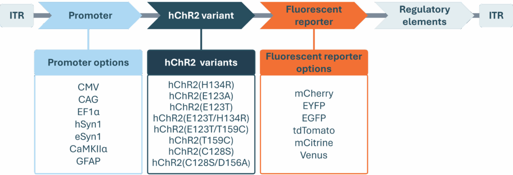
AAV opsin constructs are available with a range of commonly used promoters, including constitutive promoters (CMV, CAG, and EF1α), neuronal promoters (hSyn1, eSyn1, and CaMKIIα), and non-neuronal promoters (e.g., GFAP), allowing researchers to select an expression strategy suited to their experimental system and target cell population (Figure 2). Available fluorescent reporters include mCherry, EYFP, EGFP, tdTomato, mCitrine, and Venus, which provide options for visualization and confirmation of transgene expression. Cre-dependent configurations are also available for experiments requiring additional genetic specificity.
Together, these options allow researchers to combine the light-responsive actuator with promoter, reporter, and conditional-expression choices suited to their experimental design.
Choosing a capsid
Vector Biolabs builds its optogenetic constructs on AAV2 ITRs and packages them in commonly used AAV serotypes and engineered capsids with neuronal tropism including AAV1, AAV2, AAV5, AAVrg, AAV9, AAV-DJ, AAV-DJ8, and AAV-DJ9, along with other wild-type and synthetic capsids. Capsid choice influences cellular tropism, transduction efficiency, and spread from the injection site. AAV1, AAV2, AAV5, AAV8, AAV9, and the engineered AAV-DJ are commonly used for direct neuronal targeting, but their expression and distribution characteristics differ by brain region and experimental model. Empirical validation remains important when selecting a capsid for a specific study [17].
Highly purified and ready to use AAV vectors are supplied at client-specified titers in PBS with 5% glycerol, in volumes starting from 100 µl to 1 ml or higher. Custom optogenetic constructs ship in approximately 4–5 weeks. Pre-made control vectors are available for immediate shipment, while turnaround for other catalog and custom vectors depends on the configuration.
Celebrating a discovery that continues to transform neuroscience
The path from the phototactic behavior of Chlamydomonas to precise control of neuronal activity illustrates how fundamental biology can lead to entirely new experimental technologies. Hegemann and Nagel’s discovery of channelrhodopsins provided a remarkably simple molecular mechanism: a protein that directly converts light into ion flow. Deisseroth’s work demonstrated how that mechanism could be applied to neurons and ultimately to intact neural circuits. The Nobel Assembly describes their discoveries as laying the foundation for “a new era in neuroscience” [1].
More than two decades after the first neuronal ChR2 experiments, optogenetics continues to evolve. New constructs, targeting strategies, viral vectors, reporters, and experimental configurations are allowing researchers to ask increasingly precise questions about how neural circuits function [18].
Explore an extensive range of Vector Biolabs AAV optogenetics vectors and available ChR2 configurations for your next study, or Contact Us
References
- The Nobel Assembly at Karolinska Institutet 2026. The 2026 Nobel Prize in Physiology or Medicine. Accessed October 7, 2026
- Nagel G, Szellas T, Huhn W, Kateriya S, Adeishvili N, Berthold P, Ollig D, Hegemann P, Bamberg E. Channelrhodopsin-2, a directly light-gated cation-selective membrane channel. Proc Natl Acad Sci U S A. 2003 Nov 25;100(24):13940-5.
- Boyden ES, Zhang F, Bamberg E, Nagel G, Deisseroth K. Millisecond-timescale, genetically targeted optical control of neural activity. Nat Neurosci. 2005 Sep;8(9):1263-8. doi: 10.1038/nn1525.
- Aravanis AM, Wang LP, Zhang F, Meltzer LA, Mogri MZ, Schneider MB, Deisseroth K. An optical neural interface: in vivo control of rodent motor cortex with integrated fiberoptic and optogenetic technology. J Neural Eng. 2007 Sep;4(3):S143-56.
- Deisseroth K. Optogenetics: 10 years of microbial opsins in neuroscience. Nat Neurosci. 2015 Sep;18(9):1213-25.
- Bi A, Cui J, Ma YP, Olshevskaya E, Pu M, Dizhoor AM, Pan ZH. Ectopic expression of a microbial-type rhodopsin restores visual responses in mice with photoreceptor degeneration. Neuron. 2006 Apr 6;50(1):23-33.
- Berndt A, Yizhar O, Gunaydin LA, Hegemann P, Deisseroth K. Bi-stable neural state switches. Nat Neurosci. 2009 Feb;12(2):229-34.
- Gunaydin LA, Yizhar O, Berndt A, Sohal VS, Deisseroth K, Hegemann P. Ultrafast optogenetic control. Nat Neurosci. 2010 Mar;13(3):387-92
- Berndt A, Schoenenberger P, Mattis J, Tye KM, Deisseroth K, Hegemann P, Oertner TG. High-efficiency channelrhodopsins for fast neuronal stimulation at low light levels. Proc Natl Acad Sci U S A. 2011 May 3;108(18):7595-600.
- Yizhar O, Fenno LE, Prigge M, Schneider F, Davidson TJ, O’Shea DJ, Sohal VS, Goshen I, Finkelstein J, Paz JT, Stehfest K, Fudim R, Ramakrishnan C, Huguenard JR, Hegemann P, Deisseroth K. Neocortical excitation/inhibition balance in information processing and social dysfunction. Nature. 2011 Jul 27;477(7363):171-8
- Gradinaru V, Zhang F, Ramakrishnan C, Mattis J, Prakash R, Diester I, Goshen I, Thompson KR, Deisseroth K. Molecular and cellular approaches for diversifying and extending optogenetics. Cell. 2010 Apr 2;141(1):154-165.
- Chow BY, Han X, Dobry AS, Qian X, Chuong AS, Li M, Henninger MA, Belfort GM, Lin Y, Monahan PE, Boyden ES. High-performance genetically targetable optical neural silencing by light-driven proton pumps. Nature. 2010 Jan 7;463(7277):98-102.
- Han X, Chow BY, Zhou H, Klapoetke NC, Chuong A, Rajimehr R, Yang A, Baratta MV, Winkle J, Desimone R, Boyden ES. A high-light sensitivity optical neural silencer: development and application to optogenetic control of non-human primate cortex. Front Syst Neurosci. 2011 Apr 13;5:18.
- Mattis J, Tye KM, Ferenczi EA, Ramakrishnan C, O’Shea DJ, Prakash R, Gunaydin LA, Hyun M, Fenno LE, Gradinaru V, Yizhar O, Deisseroth K. Principles for applying optogenetic tools derived from direct comparative analysis of microbial opsins. Nat Methods. 2011 Dec 18;9(2):159-72.
- Botterill JJ, Khlaifia A, Walters BJ, Brimble MA, Scharfman HE, Arruda-Carvalho M. Off-Target Expression of Cre-Dependent Adeno-Associated Viruses in Wild-Type C57BL/6J Mice. eNeuro. 2021 Nov 24;8(6):ENEURO.0363-21.2021.
- Fischer KB, Collins HK, Callaway EM. Sources of off-target expression from recombinase-dependent AAV vectors and mitigation with cross-over insensitive ATG-out vectors. Proc Natl Acad Sci U S A. 2019 Dec 26;116(52):27001-27010.
- Haery L, Deverman BE, Matho KS, Cetin A, Woodard K, Cepko C, Guerin KI, Rego MA, Ersing I, Bachle SM, Kamens J, Fan M. Adeno-Associated Virus Technologies and Methods for Targeted Neuronal Manipulation. Front Neuroanat. 2019 Nov 26;13:93.
- Lüscher C, Emiliani V, Farahany N, Gittis A, Gradinaru V, High KA, Roska B, Sahel JA, Yizhar O, Zeng H, Deisseroth K. Roadmap for direct and indirect translation of optogenetics into discoveries and therapies for humans. Nat Neurosci. 2025 Dec;28(12):2415-2431.